Keywords
Cerebral palsy, internal rotation gait, internal hip rotation
Cerebral palsy, internal rotation gait, internal hip rotation
In this revised version of the manuscript he following changes were incorporated-
-All minor typographical errors have been corrected.
-A figure (Figure 2) has been added to document the temporary improvement in hip rotation following derotation surgery in both Patients.
-References for the modified Oxford Grading of muscle strength have been included.
-It has been made more obvious through-out the manuscript that this is a case report of two similarly presenting patients rather than a formal study. This has led to more formal analysis of recurrence of internal hip rotation in our center, focusing on the kinematic pattern of hip rotation pre and post operatively as seen in the two cases presented here.
See the authors' detailed response to the review by Mauro César de Morais Filho
See the authors' detailed response to the review by Damien Bennett
Cerebral Palsy (CP) is the most common cause of motor deficiency in young children occurring in 2.1 per 1000 live births (Oskoui et al., 2013). Internal hip rotation gait (IHRG) is common with a reported prevalence of 31.6% in bilateral CP and is a unilateral issue in the majority (78.4%) of cases (O'Sullivan et al., 2006).
IHRG in CP has been attributed to a variety of impairments associated with CP, including hip flexor, hamstring, adductor or gluteus medius tightness; femoral anteversion and hip abductor lever arm dysfunction (Arnold et al., 1997; Arnold et al., 2000; Arnold & Delp, 2001; Delp et al., 1999; O'Sullivan et al., 2006). While the correlation between static measure of femoral anterversion and hip rotation during gait is low (Braatz et al., 2013; Lee et al., 2013), femoral derotation osteotomy (FDRO) remains the ‘gold-standard’ treatment for IHRG (Niklasch et al., 2015a; Schwartz et al., 2014). This is largely a successful intervention and a recent systematic review and meta-analysis has confirmed the positive effects of this surgery on the hip and pelvis during gait (Carty et al., 2014) with long-term benefits reported up to 9 years post-surgery (Dreher et al., 2012).
However, recurrence rates of 15% to 41% have been reported (Church et al., 2017; Niklasch et al., 2015a) and in clinical practice these patients present a significant challenge. Recurrence of IHRG following surgery can be frustrating, presenting a dilemma for both therapist and surgeon regarding how best to preserve the effect of initial surgery or whether to consider repeat FDRO following recurrence. Being able to identify those patients likely to revert to internal hip rotation following FDRO would be of significant benefit to facilitate more informed surgical planning. If the decision is made to proceed with the FDRO the realistic potential for recurrence should be discussed with the family to manage expectations and potentially plan appropriate post-operative strategies to try and best preserve the effect of derotation.
Little is known about risk factors for recurrent FDRO and to our knowledge only two studies have reported on this. Church et al. (Church et al., 2017) found that those more likely to recur had slower gait velocity and higher levels of spasticity. However, this is not particularly specific as a number of factors can influence gait speed. Niklasch et al. (2015b) reported more specific risk factors for recurrence including younger age (<10 years old), reduced hip joint impulse and increased ankle plantar-flexion and internal foot progression pre-operatively.
The purpose of this clinical case report is to highlight similarities in a recurring internal hip rotation kinematic pattern in two cerebral palsy patients despite surgical intervention(s).
We analysed two patients with bilateral spastic cerebral palsy presenting with recurrent unilateral IHRG. Both patients were GMFCS level II meaning they could ambulate independently without assistive devices. The parents of both patients were seeking advice on possible repeat FDRO after IHRG recurred following previous intervention(s). The primary goal of any further surgical intervention was to improve the internal foot progression angle and the cosmetic appearance of gait. Both patients had an initial gait analysis prior to any surgical intervention at age five and nine years respectively. The current, most recent analysis was carried out at ages 17 and 15 years respectively. Intervening analyses were carried out following any surgical intervention demonstrating initial, short-term improvement in hip rotation but these analyses are not included in this case report.
Three-dimensional kinematic and kinetic data were captured using a 4 camera Codamotion cx1 active marker system (Charnwood Dynamics, Leicestershire, UK). Kinematic data were sampled at a rate of 200 Hz while force data were captured using two Kistler force plates at a sampling rate of 400Hz. Infrared markers were placed on each participant’s lower limbs as per a modified Helen Hayes protocol (Kiernan et al., 2016). Patient A demonstrated excessive internal rotation of the right hip while the left hip was more internal in Patient B. The hip kinematic graph comparing Patient A and Patient B at initial analysis demonstrates a similar degree and pattern of excessive internal hip rotation (Figure 1). Due to young age and reduced step-lengths kinetics were not collected at the initial analysis.
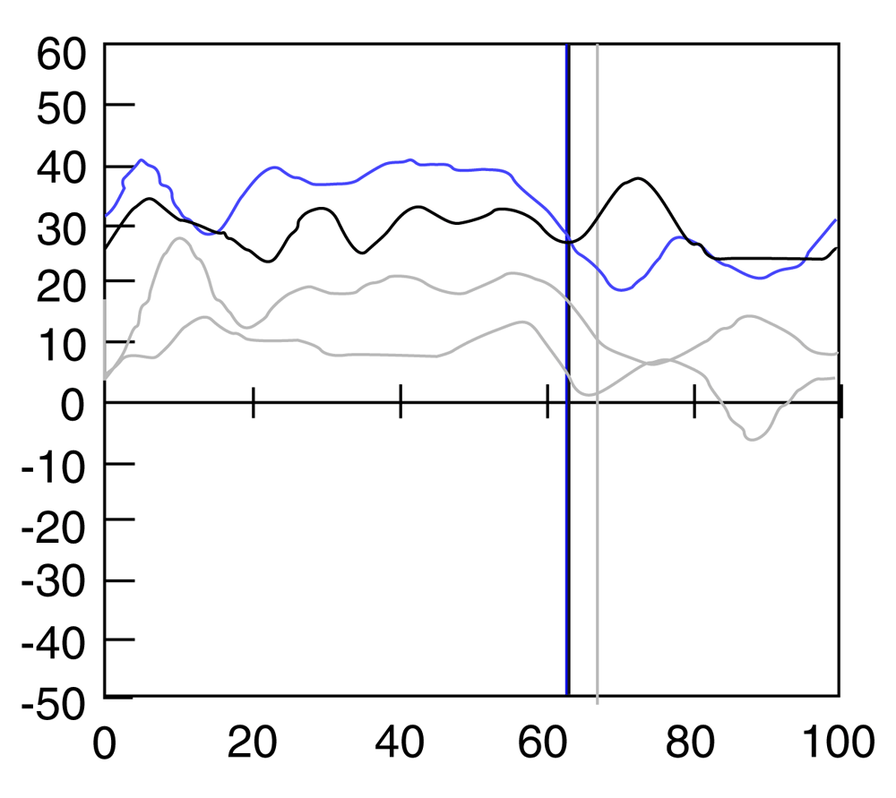
X-axis shows percent gait cycle; Y-axis shows hip rotation (internal rotation positive; external rotation negative).
Hip internal and external rotation range of movement was measured in prone lying using a gravity-reference goniometer (Myrin). Femoral anteversion was estimated in the same position using the trochanteric prominence angle test (TPAT) (Davids et al., 2002).
Hip abductor strength was assessed in side lying with the knee extended and the thigh in a neutral position in terms of flexion/extension. The limb was brought into abduction and the patient asked to hold the limb in this position while progressive manual resistance was applied. Strength was scored out of a maximum of five using the modified Oxford grading (Dan et al., 2014; Paternostro-Sluga et al., 2008). Clinical examination data at first and last assessments are summarised in Table 1.
At initial assessment, both patients demonstrated increased internal hip rotation range versus external hip rotation range and decreased strength in the hip abductors. Patient B had significantly increased femoral anteversion value at initial assessment compared to Patient A. Despite this, the dynamic hip internal rotation during gait was very similar (Figure 1) consistent with the previously reported findings that the correlation between static measure of femoral anterversion and hip rotation during gait is low (Braatz et al., 2013; Lee et al., 2013).
Patient A had a FDRO age 7 in combination with other orthopaedic procedures. The FDRO was repeated age 11 and at age 14 a surgical release of the anterior fibres of the gluteus medius was undertaken to attempt to correct the recurrent internal hip rotation. Patient B had one previous FDRO at age 11 with no additional soft-tissue releases. The current patient characteristics and past surgical histories are summarised in Table 2. In each case, post-operative gait analysis one year following intervention documented some short-term correction of IHRG but this pattern recurred in both patients (Figure 2).
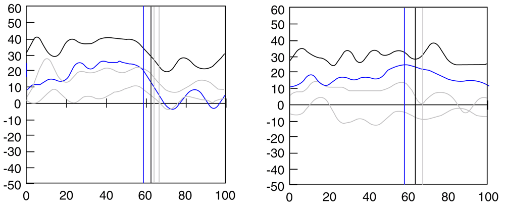
Post-operative analysis in blue; pre-operative data in black; contra-lateral pre and post-operative data in grey. X-axis of each graph shows percent gait cycle; Y-axis shows hip rotation (internal rotation positive; external rotation negative).
On review, current gait kinematics were compared to pre-operative gait patterns for both patients. In the case of Patient A there were 12 years between pre-operative analysis and current analysis and an interval of 5 years for Patient B. We found that the current post-operative degree and pattern of internal hip rotation were almost identical to their pre-operative data (Figure 3).
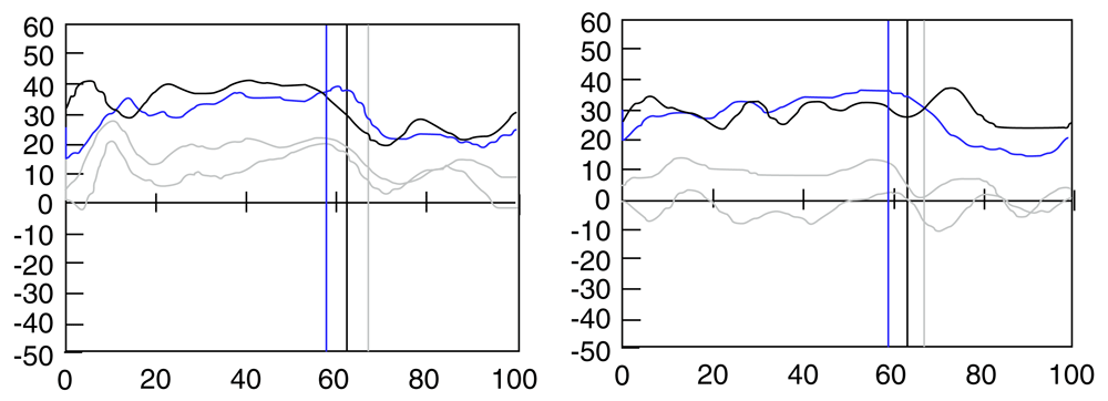
Current analysis in blue; pre-operative data in black; contra-lateral pre and post-operative data in grey. X-axis of each graph shows percent gait cycle; Y-axis shows hip rotation (internal rotation positive; external rotation negative).
On current gait analysis, both displayed some of the recently reported risk factors for recurrence of IHRG, namely reduced hip joint impulse and dynamic ankle plantar-flexion in the absence of ankle contracture. In addition, on video analysis, both had a significant trunk lean to the internally rotated side during stance indicative of probable hip abductor lever arm dysfunction.
On over-laying both patients current gait kinematics we found that both had almost identical degrees and patterns of internal hip rotation and ankle plantar-flexion during gait (Figure 4 and Table 1). This was despite otherwise different kinematics at the pelvis, hip and knee, different patient characteristics and different histories of previous surgical intervention aimed at correcting IHRG.
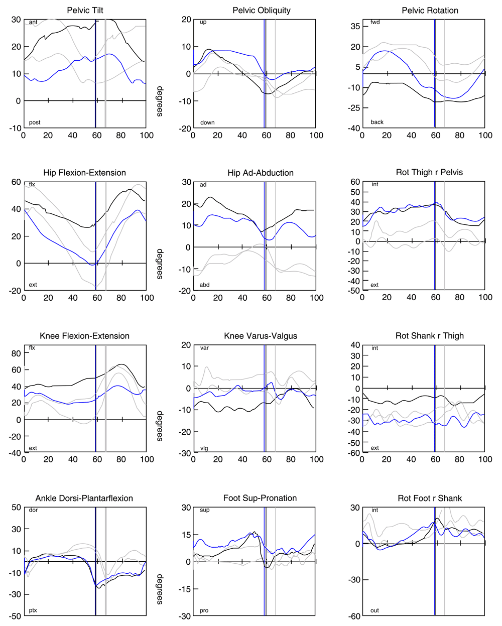
X-axis of each graph shows percent gait cycle; Y-axis shows angular displacement. Hip rotation (internal rotation positive; external rotation negative) and ankle dorsi/plantar flexion (dorsiflexion positive; plantarflexion negative) graphs circled.
The hip rotation pattern in both cases demonstrates progressive internal rotation occurring through stance phase following initial contact but a significant reduction in this internal hip rotation during swing phase. Consistent with the trunk lean and reduced hip joint impulse, this kinematic pattern further suggests that the internal hip rotation is to compensate for hip abductor dysfunction as it occurs primarily when the stance phase limb is loaded but corrects when un-loaded during swing phase.
Table 1 summarises the clinical examination findings at initial and final analysis. Despite surgical intervention, both patients continue to demonstrate significantly increased hip internal rotation versus external rotation. Femoral anteversion values have decreased compared to pre-operative values; however it must be highlighted that the reliability of the TPAT may be affected by the surgical intervention and subsequent alterations to the bony anatomy. Of note, neither patient demonstrated any change or improvement in hip abductor strength.
Our case report agrees with the findings of Niklasch (Niklasch et al., 2015b) that reduced hip joint impulse and ankle plantar-flexion during gait appear to predict recurrence of IHRG. In addition, this case report is the first report, that we are aware of, to specifically compare the joint kinematic patterns both pre and post operatively within individual patients and also between separate patients. These comparisons show that despite documented short-term improvement post-operatively after a number of surgical interventions there was a recurrence to an almost identical position of hip rotation and ankle plantar-flexion in each case. Furthermore, it appears that the hip rotation and ankle platar/dorsiflexion kinematic patterns were very similar between the two described cases.
The similarities suggest that this pattern is somehow preferential and possibly used to maximise hip abductor function. As far back as 1965, a cadaveric study found that rotation deformities of the femur represent the most efficient use of the hip abductors (Merchant, 1965). More recently, Arnold et al. (Arnold et al., 1997) used musculo-skeletal modelling to suggest that 30° of internal hip rotation best restores hip abductor moment arm. This is very similar to the recurrent position of 31–34° seen in stance phase in these two patients. The relationship between ankle equinus and hip rotation has been reported in the literature (Brunner et al., 2008). Therefore, we suggest that this position of ankle plantar-flexion during gait assists with passively internally rotating the hip.
This case report primarily presents the kinematic outcomes following surgery to address IHRG in two patients. While the similarities in hip rotation kinematics are notable, the conclusions that can be drawn from this case report are obviously limited. The findings suggest that more formal research studies on larger numbers specifically comparing kinematic patterns pre and post operatively is warranted to establish if this pattern is indeed predictive of recurrence of IHRG. Based on these preliminary findings we are now examining outcomes post FDRO in a larger group. Additionally, investigation into how this pattern develops pre-operatively is suggested as, if this internal rotation is to compensate for hip abductor dysfunction, it seems likely that growth and changes in body mass index may play a role.
In terms of our current clinical practice, the findings are consistent with previous work suggesting that in a cohort of those displaying IHRG an FDRO is not likely to offer a long-lasting solution, at least in isolation. It would appear that intervention should instead be targeted at improving hip abductor capacity but at present there is no consensus on either the cause of this hip abductor dysfunction or how best to address it. Surgery aimed at altering the pull of the hip abductor muscles has been proposed but only three studies have reported on this in CP (Cobeljić et al., 2005; Joseph, 1998; Steel, 1980) all of which have very small patient numbers and none have reported outcome measures using gait analysis. Therefore, we feel that the evidence does not currently exist for this intervention and so FDRO still offers the best potential for surgical correction of this gait pattern. However, while acknowledging the need for more formal research, we suggest that this gait kinematic pattern is a ‘red-flag’ for potential recurrence and this realistic possibility should be discussed with the relevant families. In addition, as hip abductor dysfunction is not addressed with FDRO we suggest specific focus on these muscle groups post-operatively and that a crutch or stick on the contra-lateral side be considered long-term post operatively to reduce the demand on the hip abductors and potentially reduce the need for recurrent internal hip rotation gait. Again though, future more formal research is needed to examine if these post-operative interventions preserve correction of IHRG.
Our clinical case report highlights recurrent internal hip rotation gait in two individuals with CP despite surgical intervention(s). On specifically comparing the joint kinematic graphs we have shown that in these two case reports, the position of recurrent internal hip rotation and ankle plantar-flexion are very repeatable both within each case following surgical intervention and also between the two cases. This recurrent pattern appears to be consistent with an attempt maximize hip abductor function due to the unaddressed hip abductor weakness. While the conclusions that can be drawn from this case report are limited, we now suggest that this pattern is a potential ‘red flag’ prior to surgery which should only proceed after significant discussion on potential recurrence of IHRG. As hip abductor function is not addressed with a FDRO, post-operative rehabilitation should focus on this and a stick/crutch on the contra-lateral side may reduce the demand on the hip abductors and help preserve surgical outcomes. We suggest that more formal study on the kinematic pattern in recurrent internal hip rotation is warranted and based on this preliminary case report this work is on-going in our laboratory.
Informed, signed consent for the use of anonymised gait analysis data was obtained from parents/guardians using our standard gait laboratory consent form and local institutional approval allows the use of such data.
All data underlying the results are available as part of the article and no additional source data are required.
Provide sufficient details of any financial or non-financial competing interests to enable users to assess whether your comments might lead a reasonable person to question your impartiality. Consider the following examples, but note that this is not an exhaustive list:
Sign up for content alerts and receive a weekly or monthly email with all newly published articles
Register with HRB Open Research
Already registered? Sign in
Submission to HRB Open Research is open to all HRB grantholders or people working on a HRB-funded/co-funded grant on or since 1 January 2017. Sign up for information about developments, publishing and publications from HRB Open Research.
We'll keep you updated on any major new updates to HRB Open Research
The email address should be the one you originally registered with F1000.
You registered with F1000 via Google, so we cannot reset your password.
To sign in, please click here.
If you still need help with your Google account password, please click here.
You registered with F1000 via Facebook, so we cannot reset your password.
To sign in, please click here.
If you still need help with your Facebook account password, please click here.
If your email address is registered with us, we will email you instructions to reset your password.
If you think you should have received this email but it has not arrived, please check your spam filters and/or contact for further assistance.
Comments on this article Comments (0)